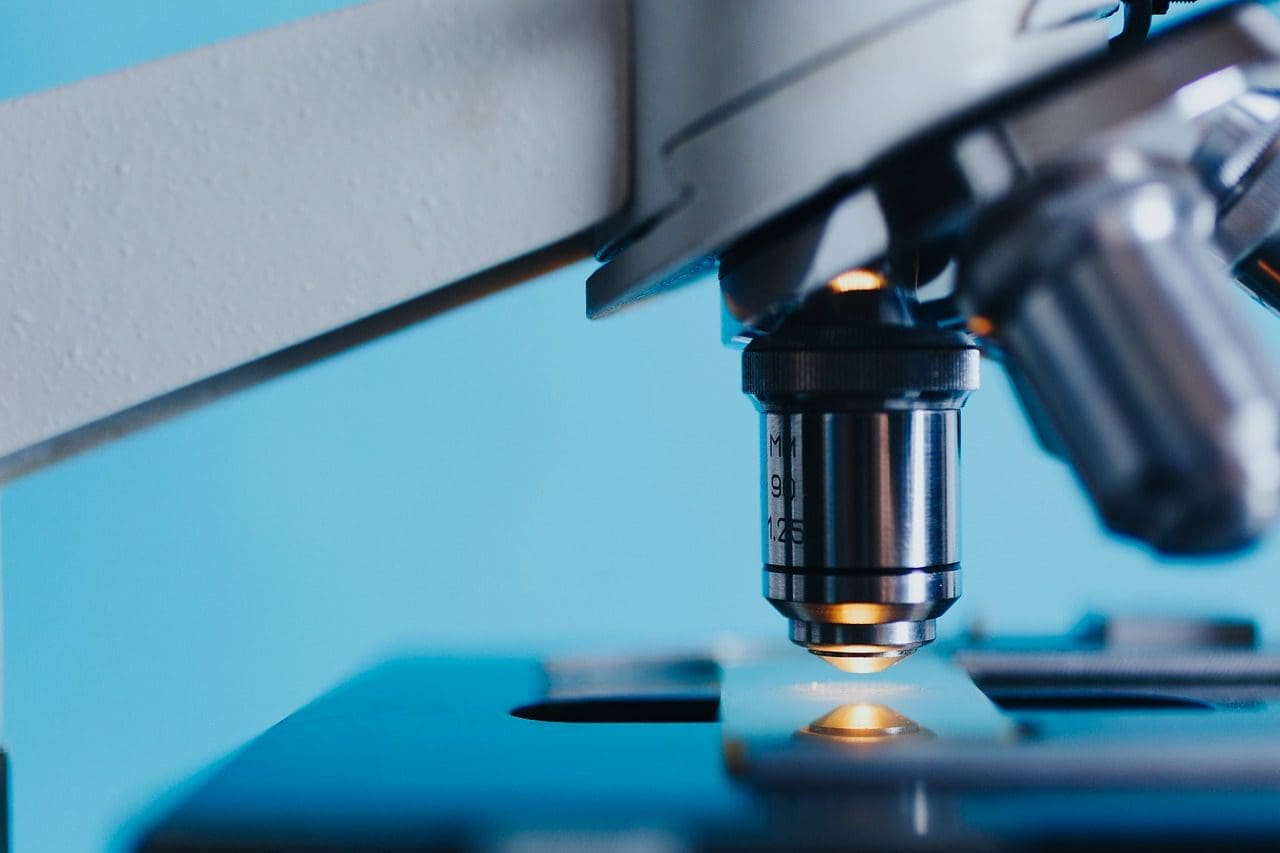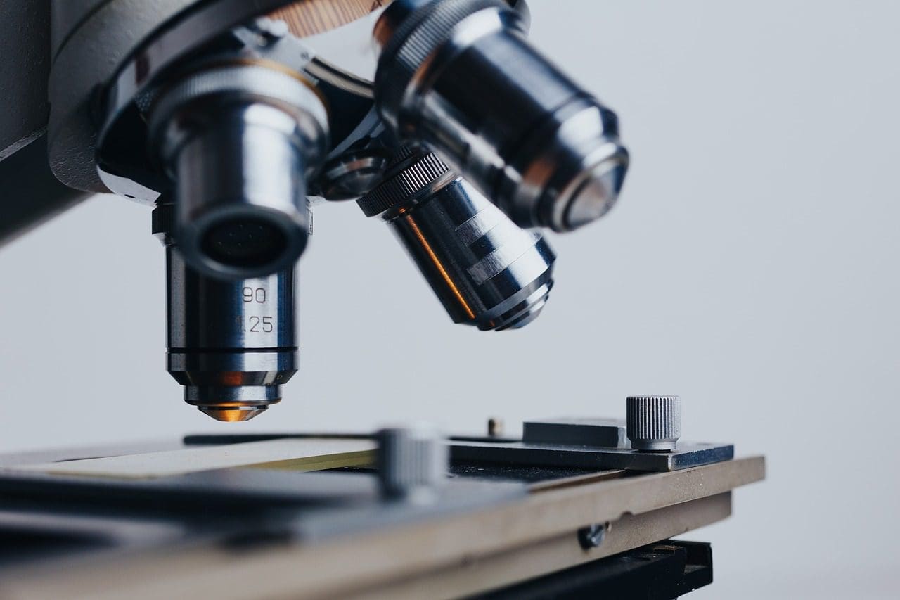
When most people think of microscopes, they think of the compound model from science class. However, there are many other types of microscopes. Researchers, medical technicians, and students utilise these gadgets regularly.
The type of microscope they choose is based on their resources and needs. Some microscopes have higher resolution at reduced magnification and vice versa and can cost tens to thousands of dollars. Therefore, let’s also distinguish between Light microscope and Electron microscope in this article.
Types of Microscopes
-
Simple Microscope
The earliest microscope is typically regarded as the simple microscope. Antony van Leeuwenhoek invented it in the 17th century, combining a convex lens with a specimen container. It was simply a magnifying glass, magnifying between 200 and 300 times. 
Despite its simplicity, this microscope was strong enough to give van Leeuwenhoek information about biological objects, including the differences in structure between red blood cells. Simple microscopes are no longer often used as the addition of a second lens led to the development of the more powerful compound microscope.
-
Stereo Microscope
The stereo microscope, often known as a dissecting microscope, can magnify objects up to 300 times their original size. Because they do not require slide preparation, these binocular microscopes are used to examine opaque objects or items that are too big to be observed with a compound microscope.
Even though their magnification is modest, they are nonetheless functional. They give a close-up, three-dimensional picture of an item’s surface textures, as well as the ability to modify the object while seeing it. Stereo microscopes are used in biological and medical research and in the electronics sector, where they are used while creating circuit boards and timepieces.
-
Compound Microscope
The compound microscope provides stronger magnification than a basic microscope because it has two lenses. The second lens multiplies the image produced by the first lens. Compound microscopes are bright-field microscopes, which means they illuminate the specimen from below, and they can be binocular or monocular.
Although the resolution is modest, these gadgets have a magnification of 1,000 times, which is considered impressive. On the other hand, this high magnification allows users to examine items that are too tiny to view with the human eye, such as individual cells. Specimens are generally tiny and transparent to some extent. Compound microscopes are utilised in a wide range of settings, from research facilities to high school biology classes, due to their low cost and great use.
-
Confocal Microscope
Unlike stereo and compound microscopes that utilise ordinary light to produce images, the confocal microscope scans coloured materials with laser light. These samples are produced on slides and inserted into the gadget, generating a magnified picture on a computer screen using a dichromatic mirror.
By combining numerous scans, operators may also produce 3-D pictures. These microscopes, like compound microscopes, have a high magnification, but their resolution is significantly superior. They’re often employed in cell biology and medical research.
-
Transmission Electron Microscope (TEM)
Like the scanning electron microscope, the transmission electron microscope (TEM) employs electrons to magnify an image. The objects are scanned in a vacuum, requiring particular preparation.
However, unlike the SEM, the TEM uses a slide preparation to produce a 2-D picture of specimens, making it more suited to studying transparent objects. A TEM is important in the physical and biological sciences, metallurgy, nanotechnology, and forensic investigation because it has a high magnification and resolution.
-
Scanning Electron Microscope (SEM)
The scanning electron microscope, or SEM, creates images by using electrons rather than light. Because samples are scanned in vacuum or near-vacuum, they must be specifically prepared through dehydration and then coating with a thin layer of a conductive substance, such as gold. 
The SEM creates a 3-D black-and-white picture on the computer screen when the item is prepped and placed in the chamber. SEMs are used by researchers in the physical, medicinal, and biological sciences to investigate a variety of specimens ranging from insects to bones because they allow them to adjust the level of magnification.
What is the difference between a light microscope and an electron microscope?
Now that we’ve seen the various types of microscopes let’s distinguish between Light microscope and Electron microscope.
A light microscope is an instrument that is used to investigate microbodies such as bacteria, fungi, and others to discover their characteristics. It also allows for the investigation of small items that are not visible to the naked eye. An optical microscope is another name for a light microscope. It is a device that enlarges pictures of microbes and other tiny creatures using light beams and lenses.
The item must be placed on a certain platform, and the microorganism must be seen via the eyepieces. Simple and compound microscopes are the two types of optical microscopes. To magnify things, the basic one makes use of the visual capacity of single or many lenses. One lens is placed near to the material to observe it in the case of complex ones.
An electron microscope, on the other hand, is a device that captures and magnifies images using electron beams. It’s a sophisticated technology that allows you to see tiny elements in great resolution. It allows you to enlarge items in nanometres and examine them thoroughly.
Conclusion
A magnifier is a piece of laboratory equipment used to magnify small objects that are too small to be seen with the naked eye. To examine and study their finer features and thus know their characteristics, we use microscopes.
The light (or optical) microscope, electron microscope, X-ray microscope, and sonic microscope are all examples of microscopes. The cell was discovered thanks to the creation of the microscope, which magnified the picture of minute things.

Be the first to comment